When examiners discover blood at a crime scene, they use a presumptive test to determine whether it is blood or not. But, to make it confirmatory, the examiner has to perform at least one of the confirmatory tests for the blood.
What are Confirmatory Tests For Blood?
To clearly depict whether the blood sample is blood or not, a special type of test is needed to perform called confirmatory test. These special tests are highly specific to blood i.e. nearly no false-positive result.
Confirmatory blood tests depends majorly on the detection of Hb molecules in RBCs. And in general scenarios, it is always done after the presumptive test for blood was performed.
Presumptive Test Vs Confirmatory Test For Blood
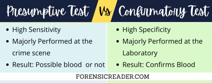
Presumptive tests are highly sensitive and less specificity to the blood. This creates a conflict about whether the bloodstain is actually blood or not. This is because the majority of the presumptive test for blood is based on the peroxidase reaction which can also be seen in some vegetables, fruits, rust, and other chemicals.
So, to come up with a surety on whether the bloodstain is actually a bloodstain or not, a confirmatory test is performed. And they are highly specific to blood.
In short, the presumptive test is highly sensitive while the confirmatory test is highly specific to blood.
| Features | Presumptive | Confirmatory |
| High in | Sensitivity | Specificity |
| Performed at | Crime scene | Laboratory |
| Result | Possible blood | Confirms Blood |
Note: You can check the dedicated article on all the 11 tests for the presumptive examination of the bloodstain.
Blood Confirmatory Test List
There are basically seven major types of confirmatory tests that can be used for the confirmation of whether the possible bloodstain is actually blood or not? These are:
- Microscopic Examination
- Intact RBCs
- WBCs Examination
- Microcrystal Assay
- Teichmann (Hematin)
- Takayama Crystal Test
- Wagenaar Test
- Spectroscopic Examination
- Electrophoresis Method
- Immunoelectrophoresis
- RNA Based Assays
- Chromatography Method
1. Microscopic Examination
Examiners first have to look for the intact RBCs. They are readily seen in cases where blood is fresh or clotted. However, when the bloodstain dries, intact RBCs are no longer visible.
Factors Affecting Microscopic Examination of Blood
Common factors are:
- Aged stains
- Environmental factors
- Heating
- Certain bleached chemicals
A. Microscopic Examination of Intact RBCs
Microscopic examination of intact RBCs is only used in fresh or clotted blood samples. As in dried stains, all cellular material is broken down and becomes unrecognizable.
Reagent Required
- Vibert’s fluid: A mixture of sodium chloride, mercuric chloride, and distilled water (or normal saline).
Procedure
- Take the stained sample and placed it inside a petri dish.
- With a dropper, add 2-3 drops (make sure stained cloth is fully soaked) of Vibert’s fluid.
- Wait for at least 6 hours, remove the stain extract, and place it on a glass slide.
- Cover the glass slide with a coverslip and observe it under a microscope.
Note: If the blood sample is in liquid form, you directly observe under a microscope. If stain is on cloth, soak the cloth sample in normal saline or Viberts fluid for at least 6 hours. However, the recommended time to be soaked is 12 hours.
Observation
- Intact RBCs confirm the presence of blood.
Species Identification From Intact RBCs
1. Human RBCs Features:
- Human RBCs are circular, biconcave, and non-nucleated.
- Average diameter of 7 μm.
- Exceptions: newborns have nucleated RBCs, and plumbism (lead poisoning).
2. Mammalian RBCs Features:
- Circular, biconcave and non-nucleated
- Exceptions: camels and yaks
3. Non-mammalian RBCs Features:
- oval, biconvex and nucleated
B. Microscopic Examination of WBCs
In some cases, looking for WBCs is known to be a viable option for the forensic examiner.
In this, staining of blood extract is done using Leishman stain. Leishman stain is used as a blood smear agent for differentiating and identifying the WBCs and malaria parasites.
So, by using this feature, WBCs can be easily identified by Leishman staining. This also elevates the chance of identifying the sex of a person by counting the Davidson bodies in polymorph cells.
2. Microcrystal Assay Test For Blood
A chemical test that results in the distinctive morphological development of microscopic crystals as a sign of blood confirmation is called the microcrystal Assay test. These microscopic heme crystals can easily be seen from a standard microscope.
Basic Principle of Microcrystal Assay Test
Altogether, they only react with the non-protein part of Hemoglobin (porphyrins) i.e.only with the oxygen-carrying protein of RBCs.
Different microcrystal assy tests produce different shaped crystals but almost react the same with Heme hexavalent.
Because of Heme’s hexavalent structure, it can interact with six different sites. Four of the heme sites are acquired by nitrogen and iron ( Fe+2) bonding. The fifth interaction is between the iron atom and the nitrogen of histidine. Lastly, either bonded with water or oxygen (oxygenated Hb).
So, if the blood is dried or made to dry (by heating), these two binding sites get active to react with different microcrystal assay reagents.
Moreover, each microcrystal test binds differently with these 2 active sites. That’s the reason why they form different microstructures as confirmatory signs of blood.
Disadvantages of Microcrystal Assay Test
It comes with majorly two disadvantages:
- It is less sensitive than presumptive test, and
- Can’t define whether the blood is human or not
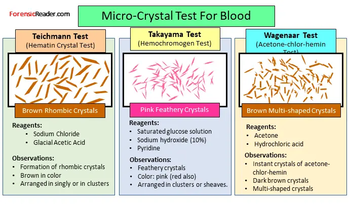
A. Teichmann Test (Hemin/Hematin Crystal Test)
The test was first documented in 1853 by Ludwik Karol Teichmann, a Polish anatomist. Other names of the Teichmann test are Hemin Crystal Assay and Hematic Crystal Assay test.
In this test, the heme part of the blood reacts with the Teichmann reagent (NaCl + glacial acetic acid) to form brown-colored rhombic crystals.
For reaction, mechanism, false positive, advantages, and disadvantages check the detailed article of the Teichmann Test.
B. Takayama Test
It was first developed by Masaeo Takayama who was a Japanese forensic pathologist in 1912. The test is used as a confirmatory test for blood with characteristic features of pink feathery crystals of hemochromogen. This is why it is also called the Hemochromogen crystal assay test.
For reaction, principle, advantages, and disadvantages, check the detailed article on Takayama Test.
C. Wagenaar Test
It is a microcrystal test where blood detection is based on the formation of dark brown crystals called acetone-chlor-hemin crystals. It was first developed in 1935 by Wagenaar.
You can get the reaction, procedure, reagent, advantages, and disadvantages from the detailed article on the Wagenaar microcrystal test.
3. Spectroscopic Examination of Blood
Spectroscopic examination of blood (or precisely hemoglobin and its derivative) is considered to be one of the most reliable confirmatory tests for both fresh and dried bloodstains. In addition, the analysis is also non-destructive in nature.
Procedure
- Take about 0.1 mg of blood in a test tube.
- Dilute the blood sample with normal saline.
- Pour a small amount of sample into a cuvette and place it in the spectrometer.
Observation
- Characteristic dark absorption bands that differ as per Hemoglobin and its derivatives.
Characteristic Spectra for Hemoglobin and its Derivatives
All the lines that are listed in the following hemoglobin derivatives are Fraunhofer lines. These are a set of spectral lines extended to the full spectrum that are used as the metric to categorize the different absorbed spectra.
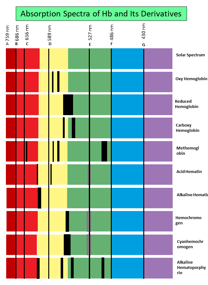
A. Oxyhemoglobin:
- Two distinct bands in yellow, between Fraunhofer lines D and E.
- One band is near D and half the breath of the other and more defined.
B. Reduced Hemoglobin:
- Broad band which lies between D and E.
C. Carboxyhemoglobin:
- Similar to Oxyhemoglobin and remains unchanged even Oxyhemoglobin gets reduced by adding ammonium sulfide.
D. Methemoglobin:
- Two similar bands are of Oxyhemoglobin with two extra bands.
- The third dark band in the red region between the Fraunhofer lines C and D.
- Fourth-one, lies between E and F which is more indistinct.
E. Acid Hematin:
- Sharp band between C and D
- Broad band between D and F, and not well-defined.
F. Alkaline Hematin:
- One band between C and D
G. Reduced Alkaline Hematin (Hemochromogen)
- One dense narrow band midway between D and E
- Second, broad and pale band over E
H. Cyan Hemochromogen
- Similar bands to hemochromogen but slightly wider.
I. Acid Hematoporphyrin
- Sharp, broad, and dark band between D and E.
J. Alkaline Hematoporphyrin
- One band between E and F broadest
- Two bands are in between D and E
- Fourth between C and D
| S.No. | Compound | No. of Bands | Fraunhofer lines (Between) |
| 1 | Oxyhemoglobin | 2 | 1st: D and E, 2nd: D and E, but close to D |
| 2 | Reduced Hemoglobin | 1 | D and E |
| 3 | Carboxyhemoglobin | 2 | Similar to OxyHb but no change on adding a reducing agent |
| 4 | Methemoglobin | 4 | 2 similar as OxyHb 3rd: C and D, 4th: E and F |
| 5 | Acid Hematin | 2 | 1st: C and D, 2nd: D and F |
| 6 | Alkaline Hematin | 1 | C and D |
| 7 | Hemochromogen | 2 | Between D and E, One is darker and the other is pale |
| 8 | Cyan Hemochromogen | 2 | D and E but slightly wider than hemochromogen |
| 9 | Acid Hematoporphyrin | 1 | D and E |
| 10 | Alkaline Hematoporphyrin | 4 | 1st: C and D, 2nd and 3rd: D and E, 4th: E and F |
4. Electrophoresis Test for Blood
Electrophoresis can be used as the confirmatory test for blood by separating and identifying the hemoglobin in the suspected sample.
Though there are lots of easy-to-perform confirmatory tests, the main reason do electrophoresis is performed by the forensic examiner is to obtain the following details:
- People’s treatment history of certain Hb abnormal diseases
- To know whether the person has a certain genetic disease
These two reasons help the case if the suspected person has some abnormal Hemoglobin disease or genetic disease.
Principle of Electrophoresis Separation
The principle of separation of Hb is based on the conjugated properties of Hemoglobins.
In general, there are various types of hemoglobin such as A1, A2, S, C, and F, and when electromagnetic fields are applied they move at different rates. This altogether resulted in the formation of various bands.
And these bands are a sign of Hb so as the confirmation of blood.
So, for analysis, the sample is taken in an appropriate buffer pH. And based on the respective opposite charge, they moved to their respective electrodes.
- Cations move toward cathode, and
- Anions move toward the anode
This lead to separation based on different types of hemoglobins in the form of bands.
Once the separation is done, hemoglobin fractions are visualized by staining with leuco malachite green solution.
Requirements For Electrophoresis of Blood
Sample Preparation (Hemolysate)
- Fresh blood samples are mixed with EDTA.
- If the blood is in clotted form, add an equal amount of water and keep it overnight in the freezer.
- Use the supernatant for the analysis.
Electrophoresis Instrument
- Cellulose acetate electrophoresis or
- Starch gel electrophoresis
Buffer ph: Hemolysate should be run either of the electrophoresis at a pH of 8.6.
Procedure
The following is the procedure for the Starch gel electrophoresis
- Take the overnight frozen sample and place it over the supporting agar gel medium.
- Electrophoresis is carried out at a constant voltage of 150 V (approx) at a pH of 8.6.
- Once the electrophoresis is done, place the supporting agar gel in a fixative to prevent diffusion.
- Add staining media such as Leuco malachite green solution.
Observation
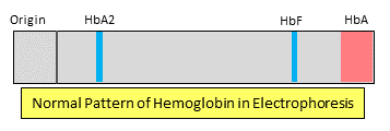
- Certain bands are formed representing different types of Hb.
- These colored bands represent the presence of hemoglobin so as the blood.
Advantages
- Very low blood sample is required.
- High separation efficiency.
- Short analysis time is required.
- Sharp band zones are obtained because of less adsorption.
Disadvantages
- Expensive than other common microcrystal confirmatory tests for blood.
- Chance of getting diffused easily.
5. Immunoelectrophoresis
In this confirmatory blood test, the identification is done on the basis of the separation of serum proteins. The term Immunoelectrophoresis is a combination of immune diffusion and electrophoresis.
This is quite a common separation and identification technique for various proteins through reaction with antibodies.
Principle
The principle of this confirmatory test depends on the first separating the antigen mixture. This can be done by the process of electrophoresis and then made to diffuse them using antisera.
So, It is a two-step process.
1st Stage: When an electric current is applied to electrodes containing agar gel, the antigen mix in the walls starts separating based on their charge and size.
2nd Stage: While the separation is done, the antigen is allowed to diffuse with antisera that are placed parallel to electrophoretic migration. These diffusions of antigen on antiserum result in the formation of distinct precipitin lines.
Procedure
- Take a small amount of bloodstain extract and place it in wells of agar on a glass slide.
- Electric current is applied to the electrophoresis rods.
- After the electrophoresis is done, the anti-human serum is placed through the length of electrophoretic migration.
- The separated protein and antiserum are made to diffuse.
Observation
- Antigen-antibody reaction with antibodies and forming precipitin lines.
- Hb has characteristically pinkish rings.
- Both are indication of blood in the sample.
Advantages
- Highly characteristically to blood detection.
- Very less sample size is required.
Disadvantages
- It takes 18 to 24 hours to get a result.
- Expensive to perform.
6. RNA Based Assays
This is not so common but majorly employed if the sample possibly contains more than one biological sample. Using RNA sequencing, the forensic examiner can look at the characteristically RNA molecules that define the origin of the sample.
So, with RNA-based assays, the serologist can then depict the origin of the fluid whether it is saliva, blood, semen, and/or their combination.
7. Chromatography Method
Chromatography can be used to confirm whether the sample has blood or not. For the most commonly used chromatography method is Ion Exchange chromatography.
Using ion-exchange chromatography, you not only separate the hemoglobin from blood (or a mixture of fluids) but also purify it.
But as per forensic importance, separating Hemoglobin from the sample is a clear sign that the crime scene sample has blood as a component.
MCQs Related to Confirmatory Test For Blood
1. Assertion (A): Confirmatory tests for blood analyses are generally spectroscopic in nature.
Reason (R): Some microcrystal tests can be confirmatory only when examined carefully by a microscopist.
- (A) and (R) both are incorrect
- (A) is correct, but (R) is incorrect
- (A) and (R) both are correct
- (A) is incorrect, but (R) is correct
2. Correct sequence of examination of an unknown bloodstain:
- Spectroscopic examination, Benzidine test, Hemochromogen test, Absorption inhibition
- Absorption inhibition, Benzidine test, Spectroscopic examination, Ring assay
- Kastle-Mayer test, Teichmann test, Oakley- Fulthrope method, mixed agglutination test
- Latte’s crust method, Absorption elution methods, Single diffusion assay, RNA-based assays
3. Arrange the following methods of blood examination in the decreasing order of examination process.
(i) Protein enzyme estimation (ii) Percipitin test (iii) Crystal test (iv) DNA analysis
- (i) (ii) (iii) (iv)
- (ii) (i) (iv) (iii)
- (iii) (ii) (iv) (i)
- (iv) (i) (ii) (iii)
4. The preliminary examination of blood can be done by:
(i) Phenolphthalein test (ii) Precepitin test (iii) Takayama Crystal test (iv) Luminal test
- (ii) and (iii) are correct
- (ii) and (i) are correct
- (i) and (iv) are correct
- (i), (ii) and (iii) are correct
5. Arrange the test methods of blood in the proper sequence:
(i) Benzidine (ii) Absorption elution (iii) Visual examination (iv) Takayama Crystal Test
- (i), (ii), (lii), (iv)
- (ii), (iii), (iv), (i)
- (iii), (i), (iv), (ii)
- (iv), (iii), (ii), (i)
References:
- Sukhadeo, et.al. Immunological Methods in Microbiology [ScienceDirect]
- Immunofixation Electrophoresis for Identification of Proteins and Specific Antibodies [PubMed]
- Development of an RNA-based screening assay for forensic [Link]
- Electrophoresis- Overview [ScienceDirect]
- Electrophoresis Principles [ScienceDirect]
- Saferstein, Criminalistics: An Introduction to Forensic Science [Book]
- Nordby, Forensic Science: An Introduction to Scientific and Investigative Techniques [Book]
- Sourcebook in Forensic Serology, Immunology, and Biochemistry [Book]
- Anil Aggrawal, Essentials of Forensic Medicine and Toxicology [Book]
- Krishan Vij, Textbook of Forensic Medicine and Toxicology [Book]
- Reddy, The Essentials of Forensic Medicine and Toxicology [Book]
Continue Reading:
- Presumptive Tests for Blood: 11 Important Forensic Test Included
- Tetramethylbenzidine (TMB) Test: Principle, Reagent and Procedure
- Forensic Analysis of Diatoms in Drowning: Extraction and Procedure
- Development of Bloody Fingerprint: Chemical Test and Techniques

FR Author Group at ForensicReader is a team of Forensic experts and scholars having B.Sc, M.Sc, or Doctorate( Ph.D.) degrees in Forensic Science. We published on topics on fingerprints, questioned documents, forensic medicine, toxicology, physical evidence, and related case studies. Know More.